

What You Need to Know About Cranio-Cervical Instability MRI
Key Takeaways
- Cranio-cervical instability (CCI) involves excessive movement between the skull and upper cervical spine, leading to various neurological symptoms.
- Accurate diagnosis of CCI often requires specialized imaging techniques, with MRI playing a pivotal role.
- Understanding the causes, symptoms, and diagnostic methods is essential for effective management and treatment of CCI.
Table of Contents
- Understanding Cranio-Cervical Instability
- Common Causes of CCI
- Symptoms Associated with CCI
- Diagnostic Imaging Techniques
- Role of MRI in CCI Diagnosis
- Treatment Options for CCI
- Importance of Early Diagnosis
- Conclusion
Understanding Cranio-Cervical Instability
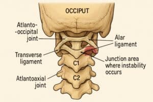
Cranio-cervical instability (CCI) is a medical condition in which the ligaments and joints between the skull and top vertebrae in the spine become loosened or weakened, leading to abnormal movement in this critically sensitive area. When movement between the skull and upper cervical spine exceeds normal limits, it can compress or irritate the brainstem, spinal cord, and associated nerves. Because the brainstem controls vital functions like breathing, balance, and heart rate, any impairment can cause significant, wide-ranging symptoms. Prompt identification and appropriate management, often involving a cervical MRI scan, are crucial for protecting neurological health and quality of life.
The cranio-cervical junction, which includes the occiput (base of the skull), the atlas (C1), and the axis (C2), is inherently more mobile than other parts of the spine due to its role in facilitating head movement. While this flexibility allows for a full range of motion, it also increases the risk of injury or instability, especially when conditions or trauma compromise ligament strength or bone integrity. Awareness of CCI among healthcare providers and patients has increased in recent years, leading to improved diagnostic strategies and more targeted treatment plans.
Those diagnosed with CCI often experience a spectrum of neurological and structural symptoms that demand individualized care approaches. Early detection through specialized imaging not only helps in confirming a diagnosis but also lays the groundwork for determining the best therapeutic options. Advances in upright and dynamic imaging technology have dramatically improved the ability to detect subtleties of instability that traditional imaging might overlook.
It is essential for individuals experiencing persistent neurological or cervical symptoms, such as unexplained dizziness, visual changes, or chronic headaches, to seek expert evaluation. Receiving the correct diagnosis is a significant step toward symptom relief and long-term management of this often-misunderstood disorder. For a deeper understanding of how neurovascular and structural disorders interconnect at the cranio-cervical junction, resources such as Johns Hopkins Medicine’s overview of atlantoaxial instability offer valuable insights.
Common Causes of CCI
- Genetic Disorders: Inherited conditions such as Ehlers-Danlos syndrome (EDS) often weaken ligaments throughout the body, contributing to laxity at the cranio-cervical junction and making these individuals more susceptible to instability.
- Trauma: Acute injuries, most notably from car accidents resulting in whiplash, as well as sports injuries and falls, can stretch or tear the ligaments holding the cervical spine in place, setting the stage for ongoing structural instability.
- Degenerative Diseases: Chronic conditions such as rheumatoid arthritis and osteoarthritis can gradually erode joints and supporting tissues, reducing the stability and alignment of the bones at the base of the skull and the upper neck.
Each of these underlying conditions increases the risk of instability and related neurological complications, underscoring the need for careful monitoring of those at heightened risk.
Symptoms Associated with CCI
The presentation of CCI can be highly variable, sometimes masquerading as other neurological or musculoskeletal disorders. Common symptoms include:
- Chronic headaches, particularly at the base of the skull
- Persistent neck pain and stiffness
- Recurring dizziness or episodes of vertigo
- Visual disturbances such as double or blurred vision
- Difficulty swallowing (dysphagia)
- Daytime fatigue and reduced concentration
Symptoms often intensify with upright posture or head movement, then improve to some extent in a reclined or lying position. This pattern is consistent with instability that worsens when gravitational forces impact the cervical spine. For some, symptoms can undermine daily functioning and overall well-being.
Diagnostic Imaging Techniques
Imaging studies are the foundation of CCI diagnosis, allowing doctors to visualize both static structures and dynamic movements at the cranio-cervical junction. Because standard imaging (like conventional X-rays and supine MRIs) may fail to identify subtle instabilities, more advanced modalities are often recommended:
- Upright MRI: Upright MRI imaging captures anatomical relationships and spinal cord compression as they occur under normal load and posture, revealing conditions that remain hidden when lying flat.
- Digital Motion X-ray (DMX): By taking rapid-sequence, real-time images, DMX visualizes the vertebrae in motion, which is essential in documenting abnormal movement patterns.
- Flexion-Extension X-rays and CT Scans: These studies require the patient to move their head forward and backward. At the same time,mages are taken to highlight the instability that surfaces only with movement.
Combining different imaging studies gives clinicians a more comprehensive view and is critical for ruling out other conditions that can mimic CCI symptoms. For those interested in further learning about the impact of spinal ligament injuries on health, the National Institutes of Health’s peer-reviewed articles on spinal instability provide an in-depth resource.
Role of MRI in CCI Diagnosis
Magnetic resonance imaging (MRI) is indispensable for evaluating CCI. Unlike X-rays and standard CT scans, MRI provides high-resolution images of soft tissues, including nerves, ligaments, and the spinal cord. This sensitivity is vital, as CCI often stems from ligamentous weakness or damage rather than bone anomalies. Upright MRI is beneficial since it closely replicates the patient’s daily experience, detecting nerve impingement and abnormal motion that static imaging cannot. Clinicians rely heavily on MRI images to assess instability severity and plan conservative or surgical interventions with precision.
Upright MRI vs. Conventional MRI
Whereas conventional MRI is performed in a horizontal position, upright MRI allows professionals to observe the cervical spine under the real-life stresses of gravity and body weight. This not only provides a more realistic assessment but also captures instability that compromises the spinal cord and neural tissue function only during regular activity. As a result, upright MRI has changed the landscape of CCI diagnosis and enables more effective, patient-centered care strategies.
Treatment Options for CCI
Managing CCI requires a multidisciplinary approach, with treatment tailored to the underlying cause and degree of instability. Non-surgical strategies are often the first line of defense, particularly in mild or moderate cases:
- Conservative Approaches: Physical therapy, cervical stabilization collars, and activity modification help limit painful movements and provide short-term support while promoting strengthening and alignment.
- Surgical Interventions: In severe or progressive cases, surgical solutions such as cervical spinal fusion may be considered. These procedures stabilize the joint, improve neurological function, and reduce symptoms, especially when conservative measures fail.
Ongoing follow-up is essential to monitor symptom progression and adjust management strategies over time.
Importance of Early Diagnosis
Detecting CCI as early as possible means clinicians can implement strategies to reduce complications, enhance quality of life, and slow disease progression. Advanced imaging, especially upright MRI, enables more accurate identification of instability and its extent, ensuring that treatment plans are precisely targeted. Early intervention is key in preventing permanent neurological deficits and can be life-changing for individuals struggling with chronic, unexplained symptoms.
Conclusion
Cranio-cervical instability is a complex but increasingly understood condition. With greater awareness among healthcare providers and patients, more accurate diagnostic imaging, and a personalized approach to treatment, outcomes for those affected by CCI continue to improve. MRI, particularly upright MRI, remains the cornerstone of diagnosis and management, empowering patients and clinicians to make informed decisions for better neurological health.